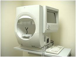
Anterior Segment Oct For Angle Study
The eventual transition from time-domain to spectral-domain OCT devices (also known as Fourier-domain OCT) has facilitated faster scanning speeds, greater tissue penetrance, and higher axial resolution images due to use of shorter wavelengths of light. Images with axial resolutions ranging from less than 5 microns (considered ultra-high resolution) to greater than 5 microns (considered high resolution) can now be obtained. Spectral domain OCT devices however have the disadvantage of having reduced scan depth compared to time domain OCT machines, due to shorter horizontal scan width.
Swept-source OCT has also been developed, enabling simultaneous acquisition of numerous longitudinal and transverse scans to create 3-dimensional corneal, anterior segment, and gonioscopy images. There are several high-quality commercially available AS-OCT machines. Some of the common spectral domain machines used include the Heidelberg Spectralis OCT, the Carl Zeiss Meditec Cirrus OCT, the Optovue RTVue and the Optovue Avanti. Common time domain machines include the Carl Zeiss Meditec Stratus OCT and Visante OCT.
Oct of Optic Nerve Head Study
How Does OCT Work? OCT is similar to an ultrasound, using light waves instead of sound waves. These light waves reflect off of different depths within the eye to reconstruct a profile, while a laterally scanning light beam creates a 3D image of the eye.
